how the body works / metabolism / nervous tissue / skeletal + circulatory / kidneys + digestion / hormones

The Skeleton provides the Framework of our Body
Skeletal Muscle Lets Us Move
The Heart and Circulation Are a Delivery System
BONE & SKELETON PROVIDE THE FRAMEWORK OF THE BODY
What is the skeleton?
The exquisite appearance of the human body is founded upon our skeleton. Our skeleton is a combination of 206 separate bones and supporting ligaments and cartilage. The bones of our skeleton are attached to muscles, which allow us to move about. Bones also provide protection. For instance, the skull and the vertebrae enclose the brain and spinal cord, respectively, thereby protecting the invaluable central nervous system (CNS).
Twelve pairs of ribs extend from our vertebrae and protect the organs of our chest. Bone also serves as a storage site for several minerals, such as calcium and phosphorus, and is the site of formation for many of our blood cells. By about six weeks of pregnancy the skeleton is rapidly developing and is visible in a sonogram. Bones continue to grow until early adulthood, complementing the growth of other body tissue. Up until this point, bones grow in both length and diameter. Around this time the longer bones of our body, such as the femur, humerus, tibia, and fibula, begin to lose the ability to grow lengthwise and our adult height is realized. Some of the bones of the lower jaw and nose continue to grow throughout our lives, although the rate of growth slows dramatically.
As you may expect, the longest, heaviest, and strongest bone in our body is the femur or thigh bone. These bones extend nearly two feet in some of us, and provide much of the support we need against the force of gravity. Meanwhile, the three small bones in the inner ear are the smallest bones in our body. In addition, the tiny pisiform bone of the wrist is also very small, having the approximate size of a pea.
What is bone?
Our fascination with the fossil remains of dinosaurs and other ancient creatures may lead us to believe that bone is a hard, nonliving part of our body and part of the bodies of other animals, including those from long ago. Although bone is indeed solid and strong, allowing form, movement, and organ protection, it is living tissue and constantly changing
Bone contains several different types of cells, which are supported by a thick fluid called the matrix. Within the matrix reside proteins, primarily collagen, and to a much lesser degree other related substances, such as some really unique carbohydrates. Also in the matrix are mineral deposits, largely a calcium- and phosphate-based crystal called hydroxyapatite, as well as calcium phosphate.
Bone is roughly 60 to 70 percent mineral complexes and the remaining bone is largely protein, primarily collagen. Hydroxyapatite are like tiny, long, and flat sheets of minerals that actually lie on top and along longer collagen fibers. These mineral deposits provide the hard and compression-resisting properties to bone. For the most part, it is also these mineral complexes along with some proteins that exist as fossils long after the death of an animal.
The bone cells (osteoblasts) in the figure above are busy making collagen proteins that form into collagen fibers that are like rope, here in the matrix of bone. Mineral complexes then adhere to the collagen. Collagen makes bone strong and minerals make it hard! In addition to some cells, proteins, carbohydrates, and minerals, other tissue can be found in bone. For instance, small blood vessels run throughout bone and deliver substances to and away from bone. Some nerves can be found in bone as well.
Is bone constantly changing?
Bone is constantly being turned over. Specific cells within bone are constantly breaking down bone components such as proteins and mineral complexes. Meanwhile, other cells are constantly building bone. Although this may seem counterproductive its merit lies in the ability of bone to adapt or be remodeled according to the demands placed upon it. For example, one of the benefits of weightlifting is an increased stress placed on bone, which causes the bone to adapt by increasing its density. In this case, the efforts of cells that build bone will exceed the efforts of cells that will break down bone components. On the contrary, prolonged exposure to zero gravity (weightlessness) in outer space will decrease the stress placed upon bone resulting in a loss of bone density. In this situation, the efforts of cells that break down bone will exceed those efforts of cells that build bone components.
SKELETAL MUSCLE ALLOWS US TO MOVE
What is skeletal muscle?
Skeletal muscle is made up of very specialized cells that have the ability to shorten when they are stimulated. With the exception of reflex mechanisms, such as the knee tap by a physician, movement of our skeletal muscle is under the command of our brain, as mentioned earlier. Because muscle cells are very long they are often referred to as muscle fiber.
The fibers are bundled up like a box of dry spaghetti or straight wires in a cable. The muscle fiber bundles are themselves bundled up and are part of larger collection of similar bundles which make up a particular muscle. Skeletal muscle is so named because it is generally anchored at both ends to different bones of our skeleton. When muscle contracts, it pulls on a specific bone, which moves the bone, thus moving a body part.
How does skeletal muscle work?
Like neurons, skeletal muscle fibers are also excitable. In fact, the excitability process of muscle cells is very similar to that of neurons, while the end result is different. Excitability in muscle fibers leads to the contraction of the muscle cell while neurons merely carry the electrical nerve impulse to another neuron or to skeletal muscle or other tissue and organs.
The inside of skeletal muscle fibers appears very different from other cells because of the contractile apparatus it contains. Each muscle fiber contains a tremendous amount of small fibrous units called myofibrils. The prefix myo refers to muscle and fibril means little fiber. Each myofibril is a stalk-like collection of proteins. The predominant proteins are actin and myosin, which are referred to as the thin and thick filaments, respectively. They are organized into a series of tiny contraction regions called a sarcomere. Myofibrils are composed of thousands of sarcomeres situated side by side.
What is calcium’s role in muscle contraction?
Calcium isn’t just important for bone, it is also the key factor that initiates muscle contraction. When skeletal muscle fibers become excited, calcium channels open and calcium floods in and around the myofibrils and bathes the sarcomeres. Calcium then interacts with specific proteins associated with actin and induces sarcomere contraction. The contraction of one muscle fiber is really the net result of the shortening of all the tiny sarcomeres in each myofibril within that cell. Further, the contraction of the muscle itself is the net result of contraction and shortening of muscle fibers that make up that muscle.
Skeletal muscle cells have another unique characteristic. They contain an organelle called the sarcoplasmic reticulum which is actually a modified version of the endoplasmic reticulum found in other cells. This organelle stores large quantities of calcium. In fact, when a skeletal muscle cell is stimulated, most of the calcium that bathes the sarcomeres actually comes from the sarcoplasmic reticulum.
What powers muscle contraction?
In order for muscle fibers to contract, a lot of ATP must be used. Some of the energy released from ATP is used to power the contraction. Interestingly, ATP is also necessary for a contracted muscle cell to “relax” as well. When the muscle is no longer being stimulated, ATP helps the thick and thin filaments to dissociate from each other so that each sarcomere can return to a relaxed (or unstimulated) position. Also, ATP is necessary to pump calcium out of intracellular fluid of the muscle fiber. Calcium is either pumped out of the cell or more likely into sarcoplasmic reticulum organelles.
If ATP is deficient, muscle fibers become locked in a contracted state called rigor. Rigor mortis occurs when the human body dies as the integrity of muscle cell membranes decrease. This allows calcium to leak into the contracting regions of muscle fibers from the extracellular fluid and from within the sarcoplasmic reticulum. As a result, calcium bathes myofibrils and contraction is invoked. Usually there is enough ATP in these dying cells to power the contraction. The dying cell would then remain locked in a contracted state.
THE HEART AND CIRCULATION ARE A DELIVERY SYSTEM
What is the heart and circulation?
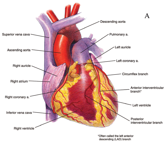
Some ancient philosophers believed that the heart was the foundation of our soul. Today we recognize the heart for its true function, that of a muscular pump. The adult heart is about the size of its carrier’s fist and weighs about one-half pound (see Heart Figure). It serves to pump blood through thousands of miles of blood vessels to all regions of our body. Blood leaves the heart through arteries on route to tissue throughout the body. Arteries feed into smaller arterioles and subsequently tiny capillaries, which then thoroughly infiltrate tissue.
Most of the miles of blood vessels is attributable to capillaries. These blood vessels are so numerous in tissue that nearly every cell in our body will have a capillary right next to it or very close. Its like having one river (artery) flowing into town, which then branches to the extent whereby every house has its own little stream (capillary).
Capillaries are the actual sites of exchange of substances between our cells and the blood. As blood reaches the end of the capillaries and the tissue has been properly served, the blood will then drain into larger venules. The venules will eventually drain into larger veins, which ultimately return blood to the heart. This is like the streams draining into larger steams, which then drain back into the larger river. Quite simply, our blood serves as a delivery system. It delivers oxygen, nutrients and other substances to cells throughout our body. At the same time, blood also serves to remove the waste products of cell metabolism such as carbon dioxide and heat from our tissue.
Our heart consists of four chambers (two atria and two ventricles), left and right. The left half, consisting of the left atrium and ventricle, serves to receive oxygen-rich blood returning from the lungs and pump it to all the tissues throughout the body. The right half of the heart, consisting of the right atrium and ventricle, serves to receive oxygen-poor blood returning from tissue throughout our body and pumps it to the lungs. Therefore, our heart functions as a relay station for moving blood throughout our body in one large loop, hence the term circulation.
How does our heart work?
Our heart is composed mostly of muscle cells that are somewhat similar to skeletal muscle cells yet retain certain fundamental differences. Although most of the events involved in contraction of heart (cardiac) muscle are the same as skeletal muscle, the heart is not attached to bone. Furthermore, our heart does not require the brain to tell it when to contract (beat). However, the brain certainly can play both a direct and indirect role in regulating the beating of our heart. The stimulus that invokes excitability in the heart comes from a specialized pacemaker region within our heart, called the sinoatrial node (SA node). The human heart may beat in excess of 2 billion times throughout a person’s life.
Unlike skeletal muscle, which pulls on bone when it contracts, the heart constricts in a wringing fashion when it contracts. As the heart contracts, the pressure of the blood inside the heart (ventricles) increases. This serves to propel blood out of the heart into the arteries. This increase in pressure also provides the driving force that forces blood to surge through our blood vessels.
What is the composition of blood?
The blood is comprised of two main parts, the hematocrit and the plasma, which can be assessed clinically. Red blood cells (RBCs) are the sole component of the hematocrit and function primarily as a shuttle for oxygen. Hematocrit is the percentage of our blood that is RBCs, which is typically 40 to 45% for an adult.
Plasma is about 55 percent of our blood. Of the plasma, about 92 percent is water while the remaining 8 percent includes over 100 different dissolved or suspended substances such as nutrients, gases, electrolytes, hormones, and proteins such as albumin and clotting factors. The remaining components of our blood are the white blood cells (WBCs) and platelets, which collectively make up about 1 percent of blood. WBCs are the principal components of the human immune system and provide a line of defense against bacteria, viruses, and other intruders. Some WBCs attack foreign invaders and useless materials while others manufacture antibodies and other immune factors. Last, but certainly not least, platelets participate in the clotting of blood.
The components of our blood. The hematocrit is comprised of RBC. Roughly 90% of the plasma is water and the remaining 10% is largely proteins, electrolytes and lipoproteins
What are red blood cells (RBCs)?
Red blood cells have the responsibility of transporting oxygen throughout the body. About 33% of the weight of a red blood cell is attributed to a specialized protein called hemoglobin. Because of this, red blood cells are often referred to as “bags of hemoglobin.” Hemoglobin is a large and complex protein that contains four atoms of iron and can bind to oxygen so that it can be transported in circulation. There are about 42 to 52 million red blood cells per cc of blood; and each red blood cell contains about 250 million hemoglobin molecules. Since each hemoglobin molecule can carry four oxygen molecules, the potential exists to transport one billion molecules of oxygen in each red blood cell.
There are two reasons for the need for such a large amount of hemoglobin in our blood. First, oxygen does not dissolve very well into our blood. Second, the demand for oxygen is extremely high in our body. Therefore, hemoglobin increases the ability of the blood to carry oxygen tremendously. Any situation that significantly decreases either the number of red blood cells or the level of hemoglobin they carry can compromise oxygen delivery to our tissues and potentially compromise function and health.
How do we bring oxygen into our body and get rid of carbon dioxide?
When the heart pumps, blood is propelled from the right ventricle into the pulmonary arteries for transport to the lungs. Pulmonary means lungs. Upon reaching the lungs and the pulmonary capillaries, carbon dioxide exits the blood and enters into the airways of our lungs. It is then removed from our body when we exhale. At the same time, oxygen enters the blood from the airways of our lungs and binds with hemoglobin within red blood cells. The oxygen-containing blood leaves the lungs and travels back to the heart as part of circulation. Thus every breath you take serves to exchange gases, bringing needed oxygen into your body while removing carbon dioxide.
How does the heart supply blood throughout our body?
As our heart contracts, blood is pumped from the left ventricle into the aorta. Blood moves from the aorta into the arteries, then arterioles, and finally tiny capillaries in our tissue. The blood leaving our left ventricle is rich with oxygen while the blood returning to our heart from tissue throughout our body has given up oxygen to working cells while acquiring carbon dioxide. This blood is then pumped by the right ventricle to the lungs to load the hemoglobin with oxygen and release carbon dioxide.
What is cardiac output?
If we were to measure the amount of blood pumped out of our heart during one heartbeat, whether it be from the left or right ventricle, we would know our stroke volume. Then, if we multiply the stroke volume by our heart rate (heartbeats per minute) we would know the cardiac output.
Cardiac Output = Stroke Volume (mL) × Heart Rate (beats/min)
Cardiac output is the volume of blood pumped out of the heart, either to the lungs or toward body tissue, in one minute. It should not matter which of the two destinations we consider, as they occur simultaneously and will have a similar stroke volume of about 5 to 6 liters (or quarts) per minute. During exercise both heart rate and stroke volume increase, which consequently increases cardiac output. For some of us, cardiac output may increase as much as five to six times during heavy exercise. This allows for more oxygen-rich blood to be delivered to working skeletal muscle
Where does the cardiac output go?
If referring to the cardiac output of the right ventricle, there is only one place for it to go: the lungs. Said another way, 100% of the cardiac output from the right ventricle is destined for our lungs. However, the blood pumped out of the left ventricle has many destinations. Under resting and comfortable environmental conditions about 13% of the left ventricle’s cardiac output goes to our brain, 4% goes to our heart, 20 to 25% goes to our kidneys, and 10% goes to our skin. The remaining cardiac output from the left ventricle (48 to 53%) will then go to the remaining tissue in our body, such as the digestive tract, liver, and pancreas.
During exercise, a greater proportion of this cardiac output is routed to working skeletal muscle. This requires some redistribution or stealing of blood routed to other less active areas at that time, such as our digestive tract. Contrarily, during a big meal and for a few hours afterward, a greater proportion of this cardiac output is routed to the digestive tract, which steals a portion of the blood directed to areas having no immediate need, such as skeletal muscle.
What is blood pressure?
Whether blood is in the heart or in blood vessels, it has a certain pressure associated with it. In fact, blood moves through circulation from an area of greater blood pressure to an area of lower blood pressure. As mentioned earlier, when the heart contracts the pressure of the blood in the ventricles increases. This establishes a blood pressure gradient that then drives the movement of blood through the blood vessels. This is somewhat like turning on a garden hose. When you turn on a garden hose, the water pressure is greatest close to the faucet (versus toward the open end of the hose). The result is that water moves from the area of greater water pressure toward the area of lesser water pressure and out the end of the hose.
We define pressure as a force exerted upon a surface and can measure it in mm Hg (mercury). If we apply this definition to our blood, we can say that blood pressure is the force exerted by blood upon the walls of a blood vessel. When blood pressure is measured two numbers are provided, for instance 120/80 or “120 over 80”. What this means is that the pressure exerted by the blood is 120 mm Hg during heart contraction and 80 mm Hg when the heart is relaxing between beats. The first number is the systolic or blood pressure when our heart contracts. The second number is the diastolic pressure and it is blood pressure when our heart is relaxing. Blood pressure is typically measured in the large artery of the arm because of its accessibility.
Skeletal + Circulatory System